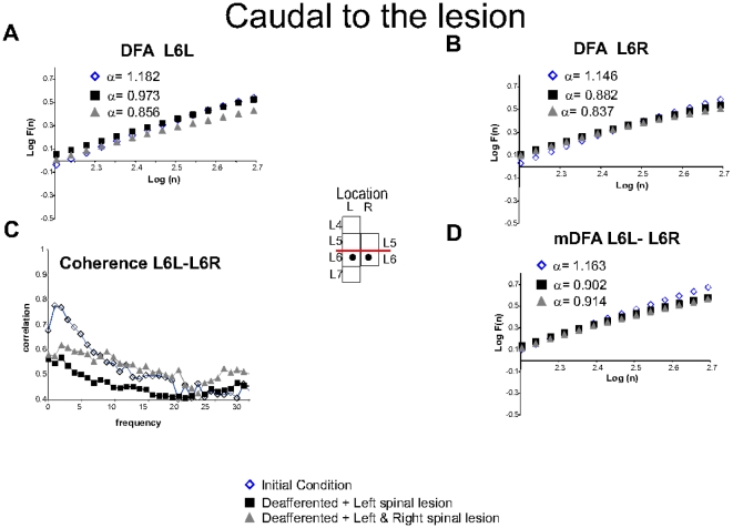
Figure 5. Spontaneous spinal recordings located below the lesion.
A. classical DFA for L6L. Insert: Location of electrodes. B. Classical DFA for L6R. C. Coherence between L6L-L6R was reduced for first lesion and followed by an increased for the second lesion as well as mDFA. D. mDFA for L6L and L6R. The regression line fit,  was above 0.99 in all cases. More details in text. Note that the spinal lesion reduced the fluctuations, but such effect was not significant.

