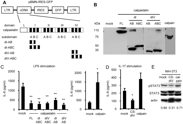
Figure 2. Retroviral expressions of calpastatin, calpastatin domains, and calpain and effect on IL-6 production from fibroblasts.
(A) Retroviral construction and scheme of the domain structure of calpastatin. Domains I and IV are abbreviated as dI and dIV, respectively. (B) Calpastatin- or calpain-GRV was transfected into phoenix 293T cells by the calcium phosphate method. Three days later, the lysates of these cells were subjected to western blot analysis. The blots were probed with anti-calpastatin and anti-calpain antibodies. (C–D) Control cells, or calpastatin- or calpain-overexpressing NIH-3T3 cells (5x10⁵ cells/ml) were cultured with 1 µg/ml LPS (C) or 10 ng/ml IL-17 (D) for 24 hours. The culture supernatants were collected and IL-6 concentrations were measured by ELISA. Left panel of (C) shows mock vs. calpastatins (Dunnett's test, n = 5), and right panel shows mock vs. calpain (Steel's test, n = 5). Panel D shows the results of three independent experiments (Steel's test). *P<0.05 and **p<0.01 vs. mock. FL, full-length. (E) Control cells, calpastatin-, and calpain-overexpressing NIH-3T3 cells (infection efficiencies were >95%) were maintained without stimulation, then lysed and subjected to western blot analysis with the indicated antibodies. Similar results were obtained in three independent experiments. Numbers below the blots are the average relative densities of pSTAT3 to actin.

