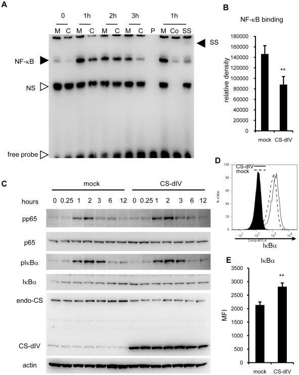
Figure 5. Overexpression of the minimal functional domain of calpastatin suppressed NF-κB signaling via stabilization of IκBα.
(A) Nuclear extracts from mock (M)- and CS-dIV (C)-overexpressing NIH-3T3 cells stimulated with LPS were subjected to EMSA. P, probe only; Co, competition by preincubation with non-labeled probe; SS, super shift by preincubation with an anti-p65 antibody; NS, non-specific bands. Similar results were obtained in six independent experiments. (B) Bands densities of NF-κB were measured and statistical analysis was performed. (C) Cytoplasmic extracts from the same cells used in panel A were subjected to western blotting with the indicated antibodies. Similar results were obtained in three independent experiments. (D) Mock- or CS-dIV-overexpressing NIH-3T3 cells were stained with IκBα antibodies intracellularly and analyzed by flow cytometry. Similar results were obtained in six independent experiments. (E) Statistical analysis of the MFI of IκBα shown in panel C. **p<0.01 vs. mock. endo-CS; endogenous calpastatin.

