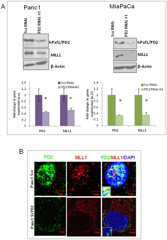
Figure 2. Expression of histone methyltransferase MLL1 in hPaf1/PD2 knockdown PC cells.
(A) Panc1 and MiaPaCa pancreatic cancer cells were transfected with scrambled and PD2 RNAi oligos and lysates were collected at 72 hours post transfection. Western Blot analysis using specific antibodies shows a decrease in MLL1 histone methyltransferase protein expression in knockdown cells compared to scrambled cells. β-actin served as a loading control. Quantification data of the western blot is provided below the corresponding immunoblot results for respective cell lines. The error bars indicated represent standard error calculated from three independent experiments. * represents significant change (p<0.05) in protein level between scrambled and PD2 knockdown cells. (B) Confocal microscope images illustrating PD2 and MLL1 localization in the nucleus of Panc1 cells. MLL1 colocalizes with PD2 in discrete regions within the nucleus (stained with DAPI) of Panc1 cells. The inset represents a magnified image of the colocalization spots of PD2 and MLL1.

