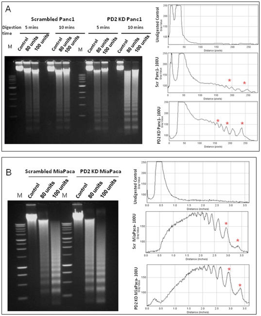
Figure 6. Micrococcal nuclease digestion altered in hPaf1/PD2 knockdown pancreatic cancer cells.
The micrococcal nuclease digestion pattern of nuclei isolated from scrambled and PD2 kd Panc1 (A) and MiaPaCa cells (B). Nuclei were digested with increasing concentrations of micrococcal nuclease enzyme (0.80 and 100 units) for 5 and 10 mins followed by DNA extraction using phenol-chloroform method. Five µg of DNA was then separated in 1.5% agarose gel. The figures on the right hand side represent densitometry analysis of the DNA banding pattern and the asterix in red shows difference between scrambled and knockdown cells.

