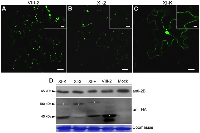
Figure 2. Transient expression of the myosin XI-K tail disrupts formation of the PD-associated tubules by the GFLV movement protein GFP:2B.
(A) Representative confocal image showing normal, PD-localized tubules formed by GFP:2B in control leaves co-infiltrated with empty vector-transformed Agrobacterium, myosin VIII-2 or XI-F tails. (B) Co-expression of the myosin XI-2 tail reduces tubules formation by GFP:2B. (C) Co-expression of the myosin XI-K tail results in the nucleo-cytoplasmic redistribution of GFP:2B; no tubules are formed under these conditions. Insets are shown in higher magnification. (D) Immunoblot analysis using 2B- (top panel) or HA-specific (middle panel) antibodies revealed similar expression levels for GFP:2B, as well as somewhat variable levels of the HA-tagged myosin tails. Bands corresponding to the class XI (100 kDa) and class VIII (40 kDa) myosin tails are marked by asterisks. Coomassie blue staining (bottom panel) shows equal loading. Scale bars, 20 µm; inside insets, 10 µm.

