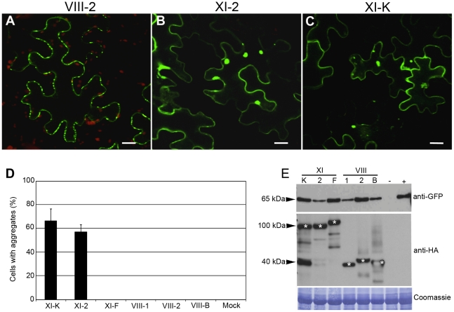
Figure 4. Transient co-expression of PDLP1:GFP and myosin XI-K or XI-2 tails leads to the PDLP1:GFP mislocalization and aggregation.
(A) PDLP1:GFP co-expressed with empty vector displays normal PD localization. Similar localization is observed in the presence of the myosin VIII-1, VIII-2, VIII-B, or XI-F tails (not shown). (B and C) Co-expression of PDLP1:GFP with the myosin XI-2 tail (B) or myosin XI-K tail (C) results in the distribution of PDLP1:GFP to the cell periphery and to formation of large cytosolic aggregates. Scale bars, 20 µm. (D) Percentage of cells showing PDLP1:GFP aggregates upon co-expression with the different myosin tail variants. Error bars indicate standard error of the mean of 3 independent experiments; between 200 and 300 cells were analysed for all experimental variant. (E) Immunoblot analysis of leaves infiltrated with PDLP1:GFP (approximately 65 kDa; top panel) and myosin tails (middle panel; marked with asterisks) as indicated. Mock-infiltrated and PDLP1:GFP controls are marked (−) and (+), respectively. The coomassie-blue stained PVDF membrane to validate equal loading is shown in bottom panel.

