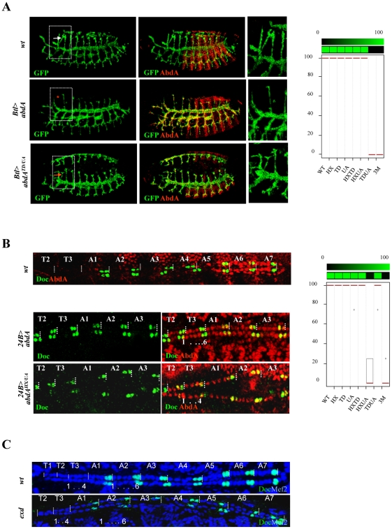
Figure 6. Functional redundancy of the HX, TD, and UA protein domains.
A. Tracheal branches are visualized by GFP-driven through the breathless btl-Gal4 driver (green). The cerebral branch (white arrowhead) forms only in T2 as a result of abdominal repression mediated by AbdA (upper panels). Expression of AbdA (red) in thoracic segments through the btl-Gal4 driver suppresses cerebral branch formation (red star, middle panels). The effect of the AbdATD/UA variants is illustrated (arrow, lower panels). Right panels are magnifications of boxed thoracic areas. B. Double immunostaining for AbdA (red) and Doc1 (green) in wild type embryo (upper panel). Magnifications of thoracic segments T2/T3 and abdominal segments A1–3 (middle and lower panels). Expression of AbdA in thoracic segments through the 24B-Gal4 driver promotes a six cell lineage state, with the two anterior most cells expressing Doc1 (middle panels). The effect of the AbdAHX/UA variants is illustrated (lower panels). Graphs in A and B (% of remaining activities compared to the wild type AbdA protein (WT) following domain mutations) using the boxplot representation summarize quantitative analyses (see Text S1 and Figure S5 (cerebral branch) and S6 (heart lineage) for full illustration). A graded color-coded bar above the graphs illustrates the level of protein activity, ranging from light green (full activity) to black (no activity). C. Abdominal hemi-segments in the cardiac tube are composed of six cardiac cells, labeled in blue by Mef2. The two most posterior cells express Doc1 (green). The thoracic hemi-segments lack the anterior Doc1 positive cells. This distinction between thoracic and abdominal segments is not affected following maternal and zygotic loss of exd.

