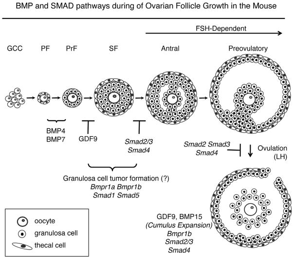
Figure 1.
Ovarian follicle development in the mouse. Oocytes are found as syncytia (germ cell cysts, GCC) in the newborn mouse ovary and GCCs breakdown to form the pool of primordial follicles (PF). Upon activation, primordial follicles transition to primary (PrF) and secondary (SF) stages. BMP4 and BMP7 are implicated in the primordial to primary transition. Follicles in mice null for Gdf9 do not progress past the primary follicle stage. In wild type mice, secondary follicles acquire a fluid-filled space (antrum) upon stimulation by FSH, and undergo cumulus expansion and ovulation under stimulation by LH. Granulosa cell-specific knockouts for Smad4 and Smad2/3 have similar defects at the terminal stages of follicle development. In contrast, granulosa cell-specific knockouts for the BMP type I receptors, Bmp1a and Bmpr1b and downstream SMAD transcription factors, Smad1 and Smad5, develop granulosa cell tumors. The follicle stage at which GCT tumors form is unknown, but based on their expression pattern [78,89], likely occurs prior to antrum formation.

