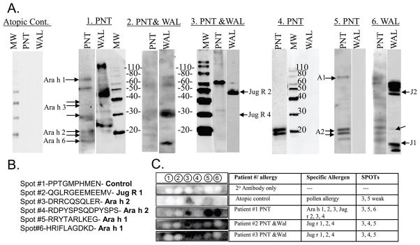
Figure 1. Immunoblots of IgE binding by peanut and or walnut allergic individuals.
A) Western blots: Patients are depicted by numbers (1–6) above each blot and the clinical allergy to peanut (PNT) or walnut (WAL). The Molecular Weight Marker (MW) is shown. Ara h 1, 2, Jug r 1, 2 and 4 are indicated as A1, A2 and J1–J4, respectively. Negative control serum is Atopic Control. B) Sequence of membrane-bound, synthetic peptides in C. C) From left to right, column 1 shows IgE binding to spots of a “no serum control” (row 1) and 4 sera to the six membrane-bound synthetic peptides (row 2–5), specific allergens recognized in western blot (column 2), and peptide spots recognized by each serum (column 3).

