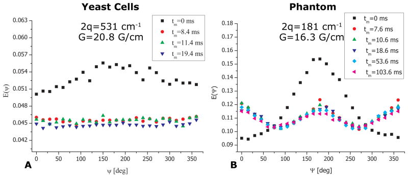
Figure 4.
bp-d-PFG experiments depicting the mixing time dependence in yeast cells and in the phantom. (A) bp-d-PFG experiments in yeast cells showing that with increasing mixing time, a flat angular dependence is observed, conveying the spherical morphology of the cells. (B) bp-d-PFG conducted in the phantom with randomly oriented cylindrical compartments with ID=29±1 μm. A qualitatively different tm-dependence can be seen, from which it can be inferred that the compartments are anisotropic, despite being completely randomly oriented. Note the pronounced modulations in E(Ψ) arising from compartment shape anisotropy.

