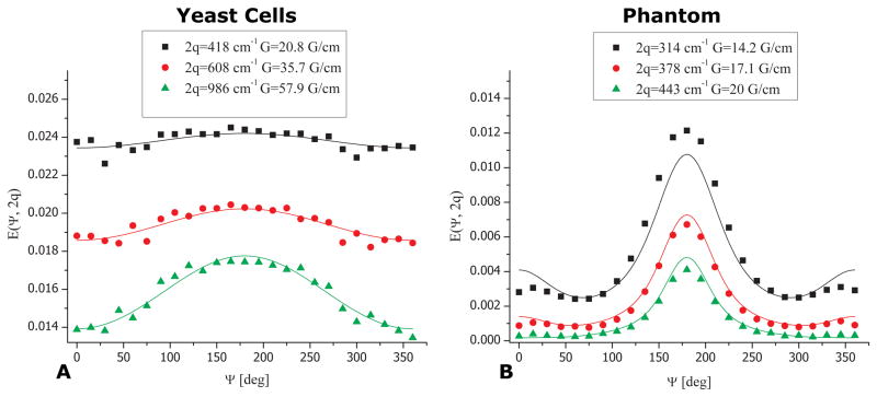
Figure 6.
Quantifying cell size from the non-invasive angular d-PFG MR. (A) d-PFG experiments (symbols) and theoretical fitting (lines) in the yeast cells show excellent agreement. The compartment size was extracted blindly and was found to be 5.46±0.45 μm. (B) bp-d-PFG experiments (symbols) and theoretical fitting (lines) in the phantom with ID=29±1 μm. The size that was extracted was 27.08±0.22 μm, in good agreement with the nominal inner diameter. The slight deviation from the nominal ID is likely due to incomplete suppression of background gradients.

