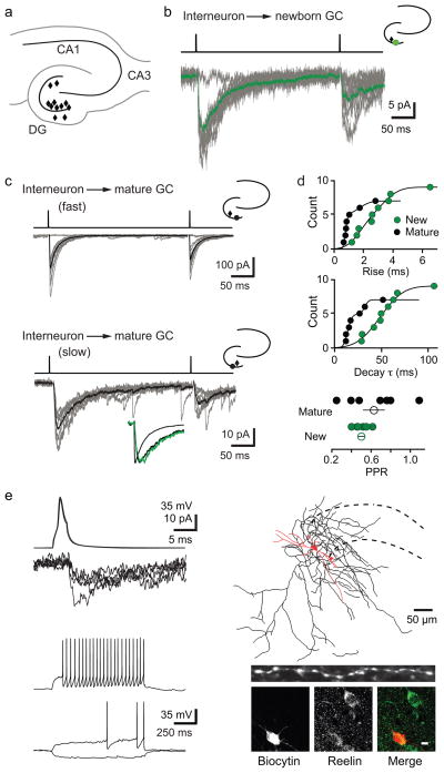
Figure 1. Ivy/NGs innervate NGCs.
a, Location of interneurons (diamonds) that innervated NGCs in acute brain slices. All procedures were approved by the UAB Institutional Animal Care and Use Committee.
b, Typical slow uPSCs in a NGC. Top, current injection protocol. Average postsynaptic response (green) is overlaid on individual uPSCs. Inset, location of pre- (black) and postsynaptic (green) cells.
c, uIPSCs in mature cells were either fast (top) or slow (bottom). Lower inset shows normalized currents from NGC (green) and mature cells (black) overlaid.
d, Rise times were fit with two Gaussian distributions with mean values of 0.78 (70%) and 1.7 ms in mature cells and 1.7 (57%) and 3.5 ms in NGCs. Decay τs were well fit with two Gaussian distributions with mean values of 14 (70%) and 32 ms in mature cells and a single distribution with a mean value of 48 ms in NGCs. PPD of uIPSCs in mature cells was more variable than in NGCs (300 ms interval). Error bars indicate S.E.M.
e, Interneuron action potentials and corresponding PSCs in a NGC (top, left) and interneuron firing pattern (bottom, left). Reconstruction of this presynaptic interneuron near the granule cell layer (dotted lines), with soma/dendrites in red (length, 820 μm) and axon in black (length, 8424 μm). Inset shows dense varicosities in a 50-μm length of axon. Post-hoc immunolabeling of reelin in the same interneuron (bottom, right). Scale bar, 10 μm.

