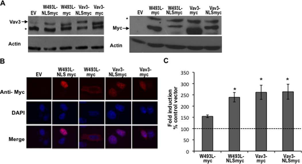
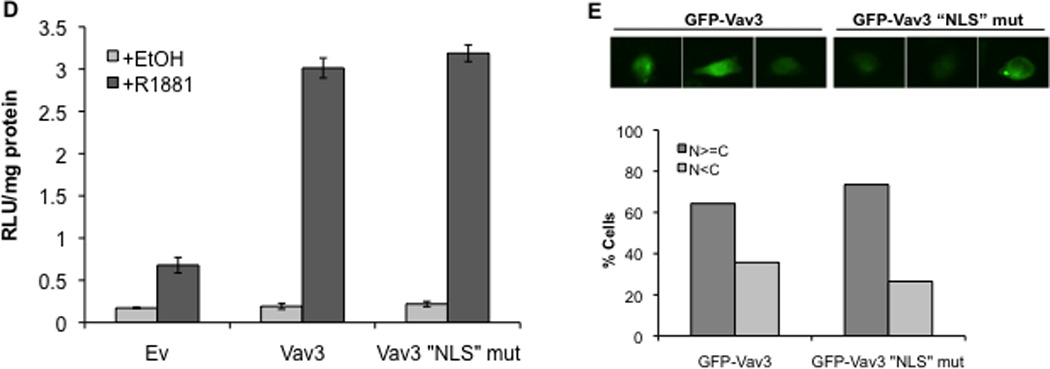
Figure 7. Nuclear targeting of Vav3 PH domain mutant (W493L) rescues AR coactivation.


A. Cell lysates from PC3 cells transfected with indicated plasmids (NLS indicates fusion with three copies of the SV40 T antigen nuclear localization sequence) were probed with anti-Vav3 (left) or anti-myc (right) antibodies. Actin was used as the loading control. A representative western blot of each is shown. Asterisks indicate non-specific bands. B. PC3 cells transfected with the indicated plasmids were stained with anti-myc (red) and DAPI (blue) and imaged by confocal microscopy. Lower images are merged with DAPI staining in blue. C. PC3 cells transfected with indicated plasmids, AR, PSA-luc and a beta-galactosidase expression vector were treated with R1881 (1 nM) or vehicle and analyzed by luciferase assay. Data (normalized to beta-galactosidase) are plotted as fold induction relative to control vector (± SEM) and represent the means from 4 independent experiments performed in triplicate. Significance was determined by two-tailed Student’s t test compared to W493L-myc (*p<0.05). D. PC3 cells transfected with AR, PSA-luc, and Vav3, Vav3 NLS mutant (Vav3NLSMUT), or empty vector (Ev) were treated with either vehicle or R1881 (1 nM) for 48 h. Cell lysate were then assayed for luciferase activity. Data from a representative experiment (of three independent experiments) performed in triplicate are plotted as relative luciferase units (mean ± SD) normalized to protein. E. PC3 cells transfected with either GFP-Vav3 or GFP-Vav3“NLS”mutant were imaged by confocal microscopy. Images of 50 cells were scored as in 6B.
