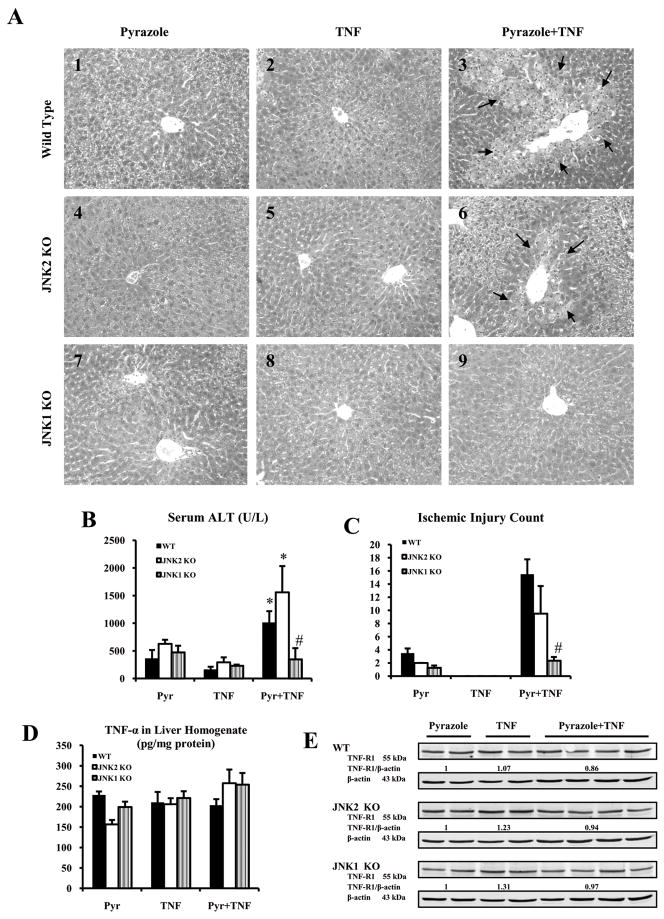
Fig.1.
Serum ALT, liver morphology and levels of TNF-α and TNFR1. (A) Histopathology. A3 and A6 show ischemic necrosis and infiltration of inflammatory cells in the central zone of the hepatic lobule (arrows, HE×300). A9 shows mild changes including hepatocyte swelling, vacuolating degeneration in the hepatic lobule. A1, A4 and A7 show mild changes including vacuolating degeneration and sinusoid congestion in the hepatic lobule. A2, A5, A8 shows no obvious pathological changes. (B) Serum ALT. (C) Ischemic injury count. (D) TNF-α in liver homogenate. (E) TNFR1 protein level.p<0.05 compared to pyrazole alone or TNF-α alone,. # p<0.05 compared to WT and JNK2 KO groups treated with pyrazole plus TNF-α.

