Fig.6.
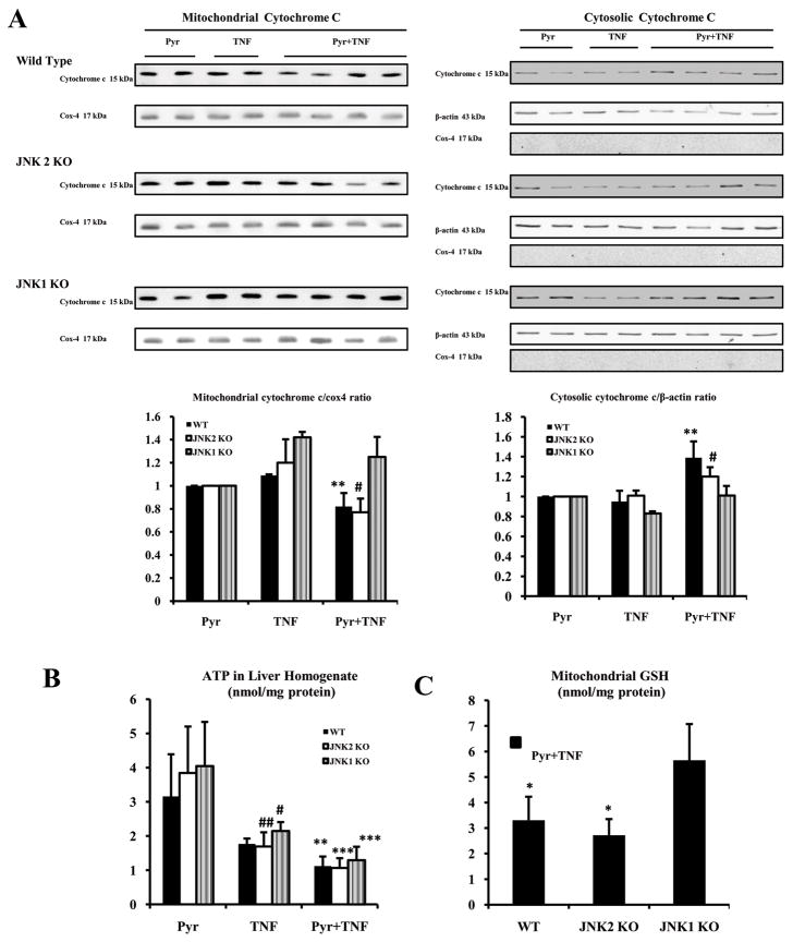
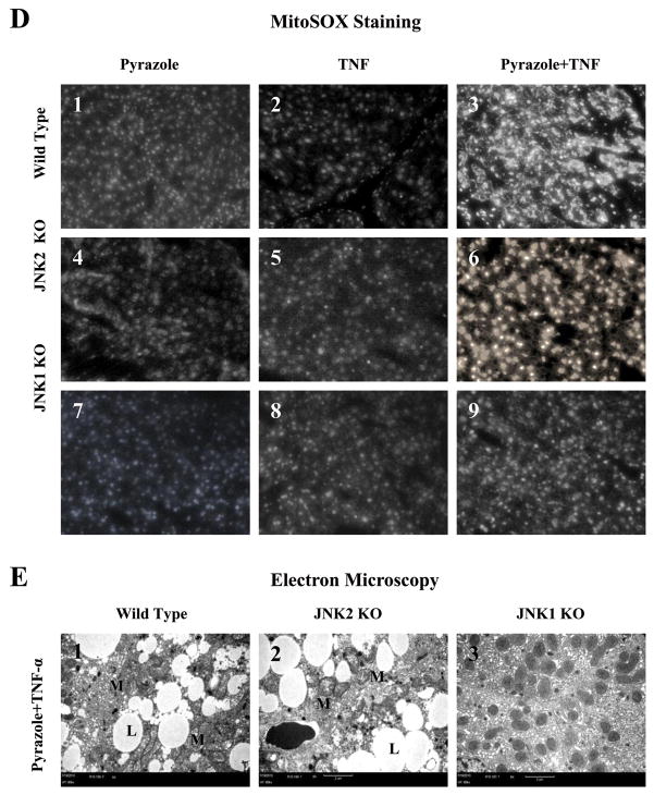
Mitochondrial assays. (A) Levels of cytochrome c protein in 20 μg of protein samples from freshly prepared cytosol and mitochondrial fractions. Cox-4 or β-actin was assayed by Western blot and results were expressed as the mitochondrial cytochrome c/cox-4 ratio or the cytosolic cytochrome c/actin ratio, # p<0.05 and ** p<0.01 compared to the pyrazole plus TNF-α-treated JNK1 KO mice. (B) ATP levels in liver homogenates were determined with recombinant firefly luciferase. ** p<0.01, *** p<0.001, # p<0.05 and ## p<0.01 compared to the pyrazole alone treatment. (C) Mitochondrial GSH. * p<0.05 compared to the JNK1 KO. (D) Fluorescence of the oxidation-dependent dye, MitoSOX Red. D3 and D6 show strong mitoSOX positive staining in hepatocytes from the WT and JNK2 KO mice treated with pyrazole plus TNF-α (+++, ×300). D9 shows weak mitoSOX positive staining in the JNK1 KO group treated with pyrazole plus TNF-α (+,×300). D1, D4 and D7(+); D2, D5 and D8 (+ or -). (E) Small liver fragments were immediately fixed in ice-cold 2% glutaraldehyde in phosphate-buffered saline and observed by transmission electron microscopy (M:mitochondria; L:lipid droplet). E1 shows severe damage of hepatocytes including lipid droplets and broken mitochondria (TEM×5000). E2 shows severe damage of hepatocyte including lipid droplets and broken mitochondria (TEM×5000). E3 shows hepatocyte with slight swelling of mitochondria (TEM×5000).


