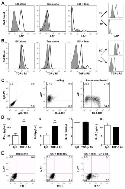
Figure 1. TGF-β inhibits IFN-γ production by alloreactive CD4+ Tem in vitro.
(A) Flow cytometric analysis of LAP surface expression on IFN-γ-pretreated EC and CD4+ Tem cultured alone or together in serum-free medium for 24 hr. Discrete cell populations from the co-cultures were also individually gated to analyze expression by EC and CD4+ Tem separately (right panels). (B) TGF-β RII surface expression on these cells. (C) LAP and HLA-DR surface expression of untreated EC placed in transwell inserts above co-cultures of CD4+ Tem with either untreated EC (resting environment) or IFN-γ-pretreated EC (immune-activated environment) for 3 d. (D) IFN-γ-pretreated EC were co-cultured with CD4+ Tem in the presence of LAP-β1 and TGF-β1/-β2/-β3 neutralizing antibodies at 10 μg/mL each or irrelevant IgG1 at 20 μg/mL for 24 hr. Cytokine supernatant levels (n=16 from 3 donors) were measured by ELISA. *P<0.05, LAP+TGF-β Ab vs. IgG, paired t test. (E) Alternatively, the co-cultured cells were incubated for a further 30 min with brefeldin A and PMA/ionomycin and then intracellular cytokine staining was performed for IFN-γ and IL-17. Dot plots show % positive cells in each quadrant and flow cytometry data are representative of 3 experiments.

