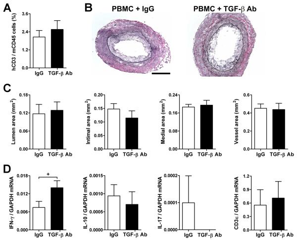
Figure 5. TGF-β inhibits IFN-γ production in early rejecting artery grafts.
SCID-beige mouse recipients of human coronary artery grafts (n=6 from 3 artery donors) received 3×10⁸ allogeneic human PBMC (from a different PBMC donor to those in Fig. 2) at 1 wk post-op and were treated with IgG or TGF-β antibody at 125 μg s.c., 3x per wk, from 1 to3 wk post-op. (A) Frequency of human CD3+ vs. mouse CD45+ circulating cells at 2 wk post-op (or 1 wk after PBMC inoculation). (B) Representative photomicrographs of EVG-stained graft sections at 3 wk post-op (bar, 300 μm). (C) Lumen, intima, media, and total vessel areas were calculated from EVG-stained graft sections. (D) IFN-γ, IL-10, IL-17, and CD3ε transcripts normalized to GAPDH were measured in grafts at 3 wk post-op by quantitative RT-PCR (IL-4 mRNA was undetectable). *P<0.05, TGF-β Ab vs. IgG, paired t test.

