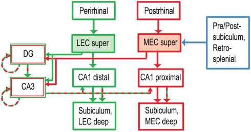
Figure 1.
Anatomical segregation of cortical inputs to hippocampus (Burwell, 2000; Witter and Amaral, 2004). The LEC receives major input from the perirhinal cortex, part of the brain’s ventral (“what”) pathway. The MEC receives input from the postrhinal (parahippocampal) cortex, part of the dorsal (“where”) pathway. The MEC also receives major spatial inputs from the presubiculum, postsubiculum, and retrosplenial cortex, all of which show stronger spatial tuning than the postrhinal cortex (Knierim, 2006). The projections of LEC and MEC to CA1 remain segregated along the transverse (proximal–distal) axis of the hippocampus, whereas the projections to the DG and CA3 converge onto the same anatomical regions. (For simplicity, a number of anatomical connections have been excluded from this diagram. super: superficial layers II and III, the inputs to the hippocampus. deep: deep layers V and VI, which receive feedback from the hippocampus.)

