Figure 5.
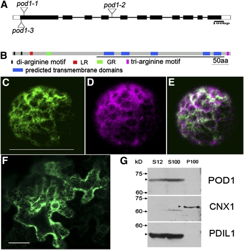
The Structure and Subcellular Localization of POD1.
(A) Three insertion alleles of pod1 designated as pod1-1 (Ds insertion line), pod1-2 (T-DNA insertion line), and pod1-3 (Ds insertion line).
(B) Domain structure of POD1 protein. The DUF747 domain is underlined. GR, Gly-rich motif; aa, amino acids.
(C) A confocal image showing POD1-GFP localization in an Arabidopsis protoplast. Bar = 50 μm.
(D) A confocal image of the same cell as (C) showing the localization of the ER marker mCherry-HDEL.
(E) Merged image of (C) and (D) shows the colocalization of POD1-GFP and mCherry-HDEL.
(F) A confocal image showing POD1-GFP localization in tobacco leaf cells. Bar = 50 μm.
(G) Immunoblot showing that POD1 protein is detected in total proteins (S12) and the soluble fraction (S100), but not the microsomal fraction (P100). CNX1 and PDIL1-1 are the controls as the membrane protein and soluble protein, respectively. Markers of molecular weight and POD1 protein are indicated on the left with arrows and arrowheads, respectively.

