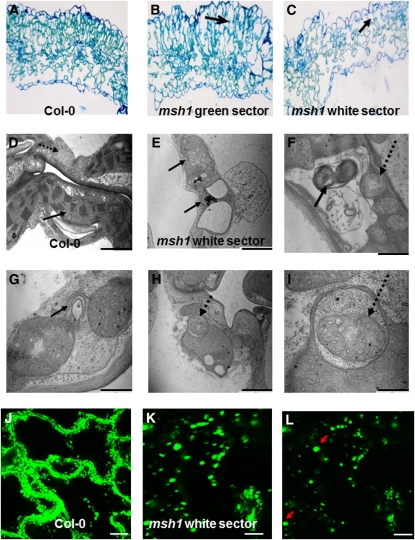
Figure 5.
Cellular and Organelle Morphology and Physiological Changes Associated with MSH1 Mutation.
(A) to (C) Cytological effects in the msh1 mutant are shown by light microscopy of a leaf cross section from wild-type (A) and green (B) or white (C) msh1 sectors. Arrows indicate loss of palisade cell organization.
(D) and (E) Transmission electron micrographs of wild-type and msh1 mutant organelle morphology in the white sector. Solid arrows indicate plastids; dashed arrows indicate mitochondria.
(F) to (I) Transmission electron micrographs show evidence of mitophagy and autophagy-like activity within the msh1 mutant.
(J) to (L) Confocal laser scanning microscopy of mitochondrial morphology and movement, visualized in wild-type and msh1 mutant plants transformed with a mitochondrial-targeting GFP construct. Time projection of mitochondrial movement over 5 min allows visualization of relative changes in mitochondrial motility ([J] and [K]). Mitochondria in msh1 white sectors are nearly stationary and enlarged. Arrows indicate unusually large-sized mitochondrial forms (single image, no time lapse) in (L). Bar = 10 μm.

