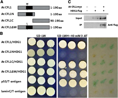
Figure 8.
Interaction between At CFL1 and HDG1.
(A) Schematic representation of the peptides used for yeast two-hybrid interaction assay. From top to bottom, the diagram represents the full sequence of At CFL1, N-terminal peptide of At CFL1, C-terminal peptide of At CFL1, and full sequence of At CFL1 with one amino acid (aa) mutated from Trp (W) to Leu (L).
(B) Yeast two-hybrid interaction analysis of At CFL1, At CFL1N, At CFL1C, and At CFL1ΔW with HDG1. Transformed yeast strains were plated on SD-LW and SD-LWH medium and colony-lift filter assays performed with X-gal, from left to right, respectively. Interaction between p53 and T-antigen was used as a positive control and interaction between lamin C and T-antigen as a negative control.
(C) Immunoprecipitation assay between Arabidopsis HDG1 and At CFL1 in vivo. Lane 1, no input; lane 2, HDG1:flag; lane 3, At CFL1:myc and HDG1:flag.

