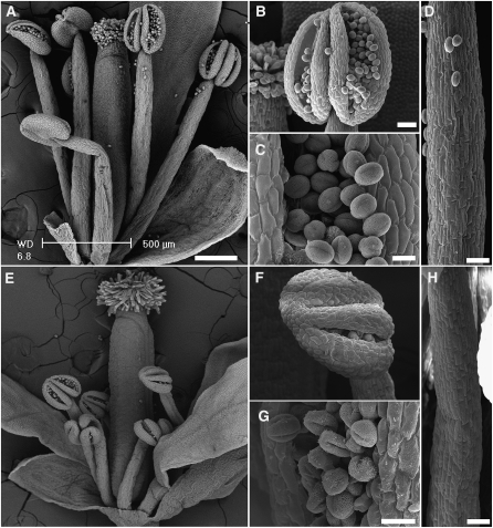
Figure 4.
Scanning Electron Micrographs Comparing the Anatomy of the Wild-Type and nhx1 nhx2 Flowers.
(A) to (D) Flowers and organs of the wild type.
(E) to (F) Flowers and organs of nhx1 nhx2.
(A) and (E) Whole flowers; (B) and (F) dehiscent anthers; (C) and (G) pollen grains; (D) and (H) filaments.
Bars in (A) and (E) = 200 μm; bars in (B), (F), (D), and (H) = 50 μm; bars in (C) and (G) = 10 μm.

