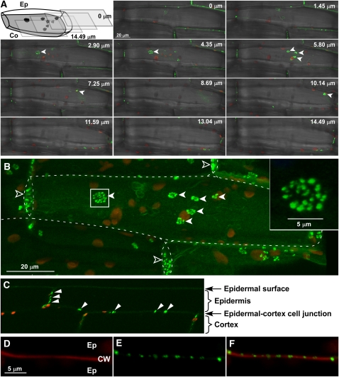
Figure 2.
PDLP5-GFP Localizes to PD Pit Fields.
Confocal images of PDLP5-GFP showing its association with PD pit fields. Closed arrowheads, PD pit fields; open arrowheads, cross-walls between epidermal cells.
(A) Confocal images shown in z-series optical sections, which span through a 14.49-μm-thick region of hypocotyl tissue across lateral wall junctions between epidermal (Ep) and cortex (Co) cells. Images show merged green, red, and transmitted channels. Illustration is included to help visualize the orientation of z-sections.
(B) A 3D maximum intensity projection reconstructed from the z-series displayed in (A). Images show merged green and red channels. Dashed lines, contour of epidermal cells; inset, high magnification of the boxed region.
(C) A reconstructed confocal image illustrating a longitudinal view of the hypocotyl tissue. Punctate PDLP5-GFP signals were only found in cross-walls. Images show merged green and red channels. Arrowheads, PD pit fields labeled by PDLP5-GFP.
(D) to (F) Confocal images showing punctate PDLP5-GFP signals within propidium iodide–stained cross-wall between epidermal hypocotyl cells, presented in red (D), green (E), and merged (F) channels. CW, cell wall.
[See online article for color version of this figure.]

