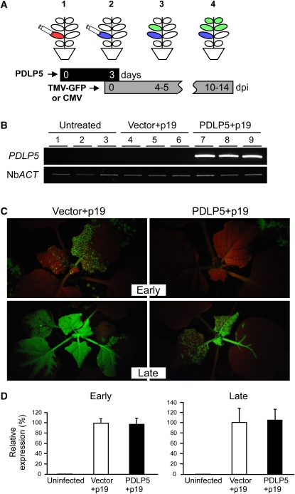
Figure 7.
Ectopic Expression of PDLP5 Delays Systemic Movement of TMV-GFP in N. benthamiana.
(A) A schematic illustration showing the experimental setup for agro-mediated PDLP5 expression and viral infection: (1) agroinfiltration of PDLP5 or empty vector control at experimental day 0 into 4th and 5th leaves (red); (2) agroinfection of the leaves (blue with TMV-GFP [or CMV inoculation] 3 d after primary agroinfiltration; (3) movement of TMV-GFP (or CMV) in systemic leaves (green) at early time points (4 to 5 DAI [dpi]); (4) systemic movement of TMV-GFP (or CMV) at late time points (10 to 14 DAI). Black and gray bars indicate the time frames for PDLP5 expression and TMV-GFP infection, respectively.
(B) RT-PCR analysis confirming the expression of PDLP5 in the leaves agroinfiltrated with GV3101 mixture carrying 35S:PDLP5 and p19 at the time of viral infection. PCR products were electrophoresed in ethidium bromide–containing 0.8% agarose gel and visualized under UV. Lanes 1 to 3, three independent plant samples that were untreated; lanes 4 to 6, three independent plant samples that were infiltrated with GV3101 mixture carrying empty vector and p19; lanes 7 to 9, three independent plant samples that were infiltrated with GV3101 mixture carrying 35S:PDLP5, and p19. N. benthamiana ACTIN (Nb ACT) was used as control for RT-PCR.
(C) Systemic TMV-GFP infection detected by GFP fluorescence under UV illumination using a Black Ray UV lamp. Representative images are shown from at least three independent repeats using 10 plants for each treatment.
(D) Systemic CMV movement detected by RT-PCR. The expression level of CMV was normalized to Nb ACT. No statistical difference in CMV detection was found between vector- and PDLP5-infiltrated samples at either early or late infection time points. Bars indicate se.
[See online article for color version of this figure.]

