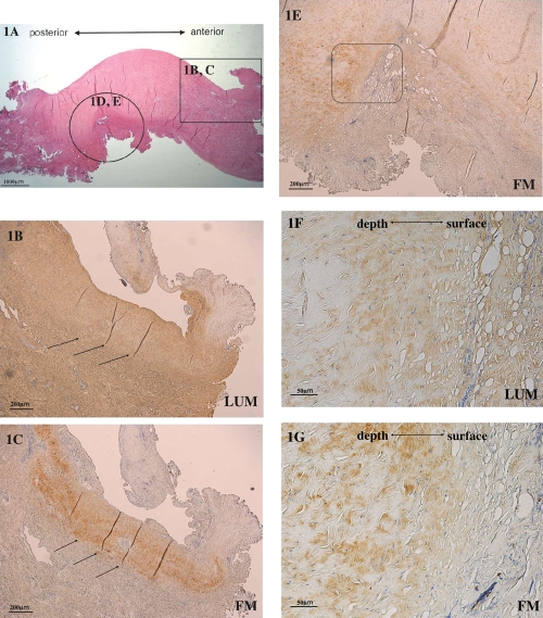
Figure 1.
Immunohistochemical analysis of Lumican (LUM) and fibromodulin (FM) expression in the deformed disc. (A) Hematoxylin and eosin stain of the deformed TMJ disc. (B) Strong lumican expression and weak lumican expression (arrows) in the deformed part of the disc (enlargement of the circle in Figure 1 A). (C) Localized fibro-modulin expression at sites of weak lumican expression (arrows) (enlargement of the rectangle in Figure 1 A). (D) Strong lumican expression in the deformed part of the disc. (E) Fibromodulin expression in the deformed part of the disc. (F) High power of the boxed region in the lumican-stained panel D. (G) High power of the boxed region in the fibrob-modulin-stained panel E (scale bars: A, 1000 µm; B–E, 200 µm; F and G, 50 µm).

