Figure 1.
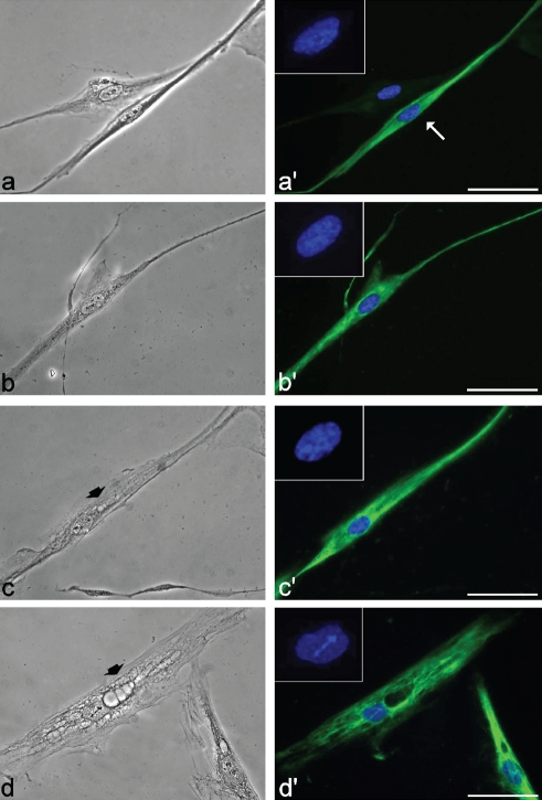
Phase contrast and fluorescence micrographs of myoblasts from healthy (a,a′ and b,b′) and DM2 patients (c,c′ and d,d′) at early (a and c) and late (b and d) passages in culture, after immunolabeling for desmin. In a′, the myoblast (arrow) may be distinguished from the fibroblast for its thin and elongated shape, and the immunopositivity for desmin (green fluorescence). Nuclear DNA was counterstained with Hoechst 33258 (blue fluorescence). Note the cytoplasmic vacuoles (arrowheads) in DM2 myoblasts (c,d). Scale bar: 20 µm.

