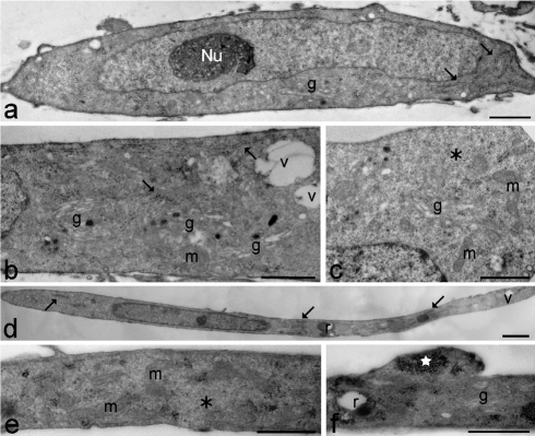
Figure 2.
Young (a–c) and senescing (d–f) healthy control myoblasts. (a) The myoblast shows an elongated shape and one ovoid nucleus with a reticular nucleolus (Nu) and scarce heterochromatin. In the cytoplasm, rough endoplasmic reticulum (arrows) and a Golgi complex (g) are visible. Scale bar: 1 µm. (b,c) Cytoplasmic details showing free ribosomes (asterisk), well developed rough endoplasmic reticulum (arrows), numerous Golgi complexes (g) and ovoid mitochondria (m). Vacuoles (v) are quite scarce. Bars: 2 µm. (d) The myoblast shows a very elongated shape and one elongated nucleus with scarce heterochromatin. In the cytoplasm, rough endoplasmic reticulum (arrows), residual bodies (r) and vacuoles (v) are visible. Scale bar: 2 µm. (e,f) Cytoplasmic details showing free ribosomes (asterisk), ovoid mitochondria (m), a Golgi complex (g), a residual body (r) and clustered glycogen granules (star). Scale bars: 1 µm.

