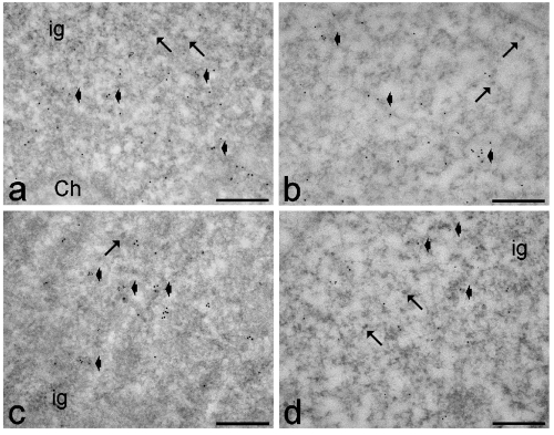
Figure 5.
Myoblasts from healthy (a,b) and DM2 (c,d) patients at early (a,c) and late (b,d) passages. Anti-polymerase II (6 nm) and anti-CStF (12 nm) antibodies: both probes specifically label perichromatin fibrils (arrowheads), while the interchromatin granules (ig) are devoid of signal. Arrows indicate perichromatin granules. Ch: bleached heterochromatin. Scale bars: 0.25 µm.

