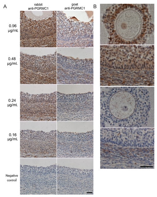
Figure 2.
Titrations of different primary antibodies to detect PGRMC1. A) Representative images of PGRMC1 localization in a medium antral follicle wall using rabbit polyclonal and goat polyclonal primary antibodies. The values on the left refer to the concentrations (µg/mL) of the primary antibodies used. Note that rabbit polyclonal antibody shows a specific staining for PGRMC1 in the granulosa cells, in the theca layer and in the endothelial cells of blood vessels at all concentration used, whereas weaker signal was detected by the goat polyclonal antibody under the same experimental conditions. No immunoreactivity was seen in negative controls where incubation with the primary antibodies was omitted. Final magnification, 200×; scale bar, 50 µm. B) Representative images showing details of PGRMC1 localization in oocytes and medium antral follicle wall using the two antibodies under the same experimental conditions at a concentration of 0.48 µg/mL. Note that rabbit polyclonal primary antibody exhibits a specific staining for PGRMC1 in cumulus cells, germinal vesicle, granulosa cells, theca layer and endothelial cells (upper images) when compared with the goat polyclonal primary antibody (lower images). Final magnification, 400×; scale bar, 50 µm.

