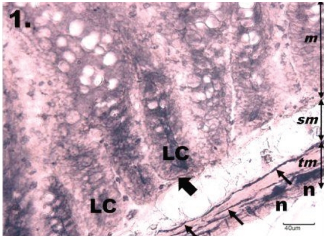
Figure 1.

Histological section through jejunum in group K. The NADPH-diaphorase positive neurons in the myenteric plexus of jejunum in the control group; tunica mucosa (m), tela submucosa (sm), tunica muscularis (tm). The myenteric plexus is intensely NADPH-d stained, neurons (n) are formed into small ganglions. Rounded or oval shaped neurons are seen located along the nerve fibres (thin arrows) running throughout the tunica muscularis. The mucosa of the jejunum is arranged into regular darkly stained intestinal glands or Lieberkühn's crypts (LC), which are bordered by NADPH-d positive nerve fibres (thick arrow). Scale bar = 40 µm.
