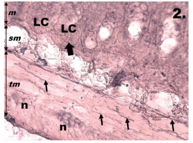
Figure 2.

Histological section through jejunum in group R1. One h reperfusion after the ischemia causes a decrease of NADPH-d positivity in the tunica muscularis of the jejunum; tunica mucosa (m) tela submucosa (sm), tunica muscularis (tm). The neurons of myenteric plexus (n) are weakly NADPH-d stained, variable in their shape, size and in the arrangement into the ganglion. The nerve fibres (thin arrows) running throughout the tunica muscularis are interrupted, the neurons are seen with unclear contours of their cell bodies, nuclei, and cytoplasmic processes. The intestinal Lieberkühn's crypts (LC) of the mucosa do not show the NADPH-d positivity, only nerve fibres bordering them are present (thick arrow). Scale bar = 40 µm.
