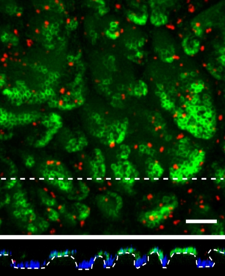
Figure 2.
Rendered confocal Z-stack of whole-mounted abdominal human epidermis stained for the stem cell marker MCSP (green), the proliferation marker, Ki67 (red) and the nuclear stain DAPI (blue). Lower panel shows the basal layer in cross sections along the dashed lines. Scale bar = 100 µm. (Online version in colour.)

