Figure. 5.
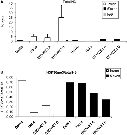
H3 occupancy (A) and H3K36me3 (B) in the intron and 5′-exon end of ERVWE1 in BeWo and HeLa cells. Using the anti-H3K36me3 and anti H3 antibodies we immunoprecipitated chromatin from BeWo and HeLa cells and analyzed the amount of immunoprecipitated intron fragment (open bars) and 5′-exon end (solid bars) by qPCR. HeLa cells were either intact or with stably integrated ectopic ERVWE1 (ERVWE1 A, B). (A) The anti-H3CT antibody was used to quantify the total H3 associated with intron and 5′-exon end of ERVWE1 as an average percentage of the input DNA. IgG non-specific controls are shown (gray bars) as an average percentage of the input DNA. Data are presented as a mean ± SD from three triplicates. (B) The enrichment for H3K36 was normalized to H3 occupancy and the ratio of H3K36me3/total H3 in intron (open bars) and 5′-exon end (solid bars) of ERVWE1 is shown.

