Figure 2.
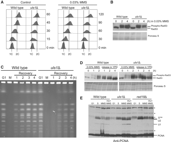
DNA synthesis defect in the uls1Δ mutant. (A) uls1Δ cells exhibit S phase delay in the presence of MMS. G1-synchronized cells of wild-type and uls1Δ were released in the presence or absence of MMS and DNA content was measured by flow cytometry at the indicated time-points. (B) Rad53 phosphorylation is increased in uls1Δ cells in response to MMS. Protein samples were prepared from G1-synchronized cells before and after release in the presence of MMS at indicated time-points and analyzed by western blotting with anti-Rad53 antibodies. (C) Completion of DNA replication is delayed in uls1Δ after MMS treatment. DNA samples from cells arrested in G1 by α factor (G1), released and treated with 0.03% MMS (M) for 60 min and then recovering in fresh YPD media were subjected to PFGE analysis. (D) DNA damage checkpoint activation does not persist in uls1Δ following MMS treatment. Protein extracts were prepared from the wild-type and uls1Δ cultures treated with 0.03% MMS for 2 h, then washed and allowed to recover in fresh media. (E) PCNA mono- and polyubiquitination is increased in the absence of Uls1. Cells were synchronized in G1, allowed to enter S phase and after 30-min incubation in YPD media exposed to 0.03% MMS or 0.03% MMS plus 0.1 M HU for 2 h. The rad18Δ mutant lacking PCNA ubiquitination was analyzed as a control. Non-specific bands are depicted by asterisks.

