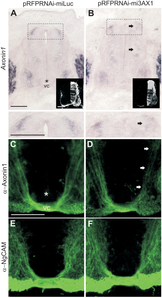
Figure 7.
miRNAs against Axonin1 (AX1) effectively and specifically knockdown Axonin-1 in vivo. Embryos were electroporated at HH17-18 with pRFPRNAi vectors driving the expression of miLuc (A, C and E) or mi3AX1 (B, D and F) and processed at HH26 for Axonin1 in situ hybridization (A and B), Axonin1 immunolabeling (C and D) or NgCAM immunolabeling (E and F). The right half of the spinal cord was targeted by electroporation, as shown by RFP expression (insets). Axonin1 is expressed by dI1 commissural axons whose cell bodies reside in the dorsal spinal cord (A, boxed region and detail). Arrows show the reduction of Axonin1 levels on the electroporated side in the presence of mi3AX1 (compare to control side). The floor-plate (asterisks) and ventral commissure (VC) are indicated. Scale bar: 100 µm.

