Figure 8.
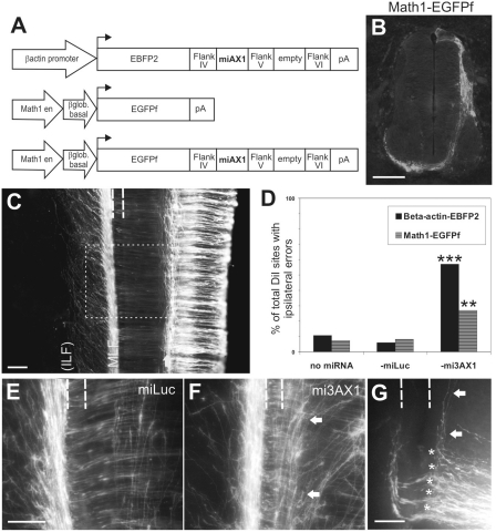
A dI1-specific miRNA construct driven by the Math1 enhancer effectively knocks down Axonin1 in vivo. (A) Schematics of the constructs. (B) Specific expression of EGFPf in dI1 axons after unilateral electroporation of Math1-EGFPf. (C) Top view of an open book taken from a HH26 embryo electroporated with Math1-EGFPf showing the trajectories of dI1 axons. A subset of dI1 axons turns ipsilaterally in the intermediate spinal cord (arrow). The majority of axons grow across the floor plate (indicated by dashed lines) before turning rostrally along the contralateral floor-plate border and extending within the MLF. The axons are later deflected diagonally and then re-converge to form the ILF (by HH27-28). The boxed area indicates the equivalent regions shown in (E and F). (D) Quantification of the phenotypes observed by DiI injection. **P < 0.001, ***P < 0.0001. (E) Visualization of fluorescent dI1 axons in embryos electroporated with Math1-EGFPf-miLuc reveals that at HH26 the majority of axons have crossed the floor plate (dashed lines) and extend rostrally along the contralateral floor-plate border. (F) Embryos electroporated with Math1-EGFPf-mi3AX1 displayed aberrant navigation of dI1 axons. Many axons were unable to enter the floor plate, instead growing longitudinally along the ipsilateral floor-plate border (arrows). (G) Visualization of commissural axons with DiI following electroporation with Math1-EGFPf-mi3AX1 confirms that the axons make ipsilateral turning (arrows) and stalling (asterisks) errors. Compare the phenotype to Figure 4H. Rostral is to the top in C, E–G. Scale bar: 100 µm in B, C, E (applies to F); 50 µm in G.

