Figure 1.
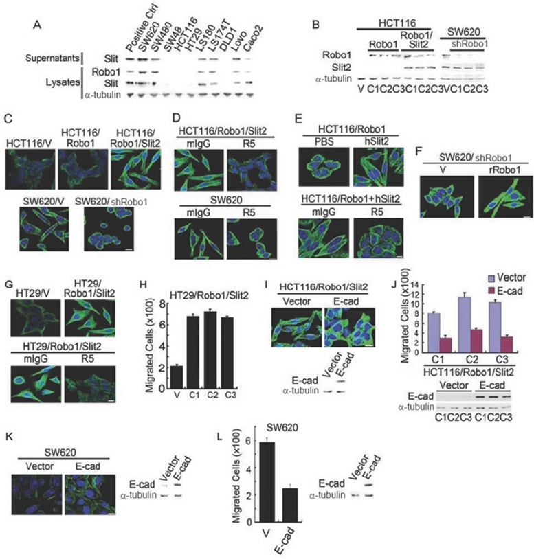
Slit-Robo signaling elicits fibroblast-like morphology. (A) Slit2 and Robo1 expression in human colorectal carcinoma cell lines detected by immunoblotting analysis with mAbs to Robo1, Slit2 and α-tubulin. (B) HCT116 cells stably transfected with plain vector (HCT116/V) or plasmid encoding Robo1 (HCT116/Robo1) or Robo1 plus Slit2 (HCT116/Robo1/Slit2) and SW620 cells stably transfected with plain vector (SW620/V) or plasmid encoding shRNA for Robo1 knockdown (SW620/shRobo1). Detergent lysates were immunoblotted with the Robo1, Slit2 and α-tubulin mAbs. C1-3, single cell clones 1-3. (C-F) Morphological features of HCT116/V, HCT116/Robo1 (C1), HCT116/Robo1/Slit2 (C1), SW620/V and SW620/shRobo1 (C1) cells in the absence and presence of mIgG, R5, PBS and hSlit2 (C-E) or transfected with the plain vector or the plasmid of rat Robo1 (F). They were immunofluorescently stained for α-tubulin (green) and nuclear DNAs with DAPI (blue). (G-L) Morphologic and migratory features of HT29/V and HT29/Robo1/Slit2 cells, HCT116/Robo1/Slit2 cells or SW620 cells transfected with the plain vector or the plasmid of E-cad in the absence and presence of mIgG or R5. Cell migratory activities were determined using the Boyden chamber assay as described previously 16. Lysates were also immunoblotted for E-cad and α-tubulin. Bars, 10 μm. Results are representative of at least three separate experiments (A-G, I and K) or the mean ± S.D. of three independent experiments (H, J and L).

