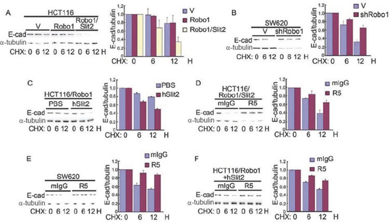
Figure 6.
Slit-Robo signaling degrades E-cad. (A-F) HCT116/V, HCT116/Robo1, HCT116/Robo1/Slit2, SW620/V and SW620/shRobo1 cells were treated with CHX, in the absence or presence of mIgG, R5, PBS and hSlit2, for a period of time as indicated. Cell lysates were immunoblotted with the Abs to E-cad (left upper panels) and α-tubulin (left lower panels) and a ratio of E-cad over α-tubulin (right panels) was calculated. Results are representative of at least three independent experiments (left panels) or the mean±S.D. of three independent experiments (right panels).

