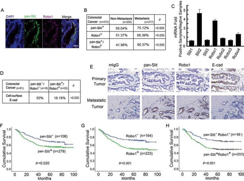
Figure 9.
Clinical significance of Slit-Robo expression in colorectal carcinoma. (A) Immunofluorescent staining of pan-Slit, Robo1 and nucleus (DAPI) and their co-localization in the colorectal carcinoma tissue specimen. Bars, 0.1 mM. (B-D) Expression of pan-Slit and Robo1 antigens (B) and Slit1-3 and Robo1-4 mRNAs (C) and their association with cell-surface E-cad (D) in the tissue specimen of non-metastatic and metastatic colorectal carcinoma. Pearson's χ2-test was used for statistical analysis (B, D). (E) Immunohistochemical staining of pan-Slit, Robo1 and E-cad in continuous sections of primary (upper panels) and metastatic (lower panels) colorectal carcinoma. Results are representative of at least 10 independent experiments. Bars, 0.1 mM. (F-H) Long-term survival curves of colorectal carcinoma patients based on the immunoreactivity of pan-Slit (F), Robo1 (G) and pan-Slit plus Robo1 (H).

