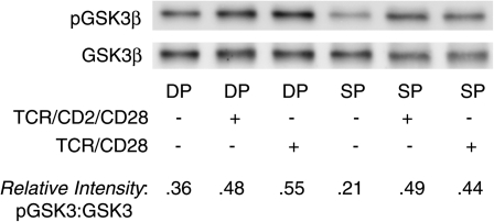
Fig. 3.
Phosphorylation of GSK3 at Ser9/21 does not differ between immature CD4+CD8+ thymocytes and mature SP T cells. Purified DP thymocytes and SP T cells were incubated at 37°C with anti-TCR/CD2 or anti-TCR/CD2/CD28-coated plates for 1 h, after which they were lysed and prepared for western blotting with anti-phospho-GSK3 (Ser9/21) and anti-total GSK3. Phospho-GSK3β shows up at ∼46 kD. (Phospho-GSK3α shows up as a much fainter band at ∼52 kD and is not always evident on western blots.)

