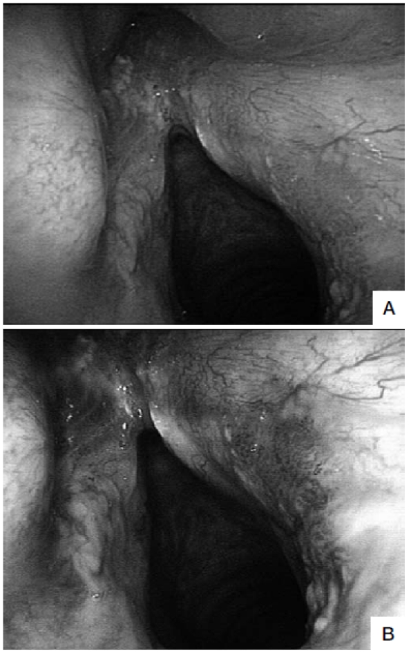
Fig. 2.

A. Follow-up examination by flexible WL endoscopy 12 months after trans-oral laser extended cordectomy (Type Va) showing a recurrent leucoplakia in the anterior commissure and a diffuse erythroplakia of the posterior third of the right vocal cord (negative at WL examination). B. The same view at flexible NBI videoendoscopy shows that the erythroplakia is a well-demarcated, brownish area with thick dark spots and an afferent hypertrophic vessel that branches out in small vascular loops in the context of the lesion (positive at NBI). Histopathological evaluation of the leukoplakia was consistent with mild dysplasia, while the erythroplakia was found to be a micro-invasive carcinoma.
