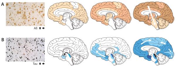
Figure 1. The accumulation of misfolded proteins in AD follows characteristic and predictable patterns.
Cross-sectional autopsy studies indicate that β-amyloid plaques (A) first appear in the neocortex, followed by the allocortex and finally subcortical regions21. In the brain, neurofibrillary tangles (B) occur first in the locus coeruleus and transentorhinal area and then spread to the amygdala and interconnected neocortical brain regions8, 24. The relatively stereotyped patterns of expansion suggest the involvement of neuronal transport mechanisms in the spread of proteopathic seeds. Increasing density of shading indicates increasing pathology. The schemata with the progression of the Aβ and tau lesions have been modified from previous publications21.

