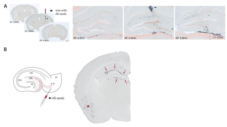
Figure 2. Induction and spread of Aβ lesions in a transgenic mouse model.
(A) Stainless-steel wire segments were coated with Aβ-rich brain extract, dried, and implanted unilaterally into the hippocampus of APP23 transgenic mice. Four months later, immunohistochemical analysis with an anti-Aβ-antibody revealed strong local induction of Aβ-deposits in the vicinity of the wire (arrowhead in the middle section of three coronal sections along the anterior-posterior (AP) axis through the hippocampus). Higher magnification of the dentate gyrus double-stained with anti-Aβ-antibody and Congo red revealed spreading of Aβ-deposition thoughout the dentate gyrus (distance between the sections shown: 600μm). Reproduced from52 with permission. (B) The injection of Aβ-rich brain extracts induces Aβ aggregation within the injected brain region, as shown here for the entorhinal cortex (EC) in APP23 transgenic mice (asterisk). However EC injections also induce β-amyloid deposition (arrows) in the outer molecular layer (OML) of the hippocampal dentate gyrus (DG), a region that is non-contiguous but is axonally interconnected with the injection site. For details of the methods, see52.

