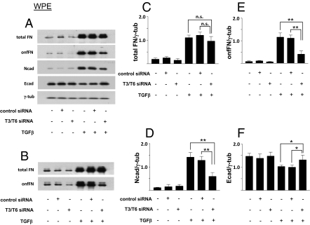
Fig. 5.
Effect of knockdown of GalNAc-T3/T6 on TGF-β–induced EMT in WPE cells, assessed by expression of epithelial and mesenchymal cell markers. WPE cells were transfected with siRNA duplexes or negative siRNA, and treated with TGF-β, as in Fig. 4. Cell lysates were prepared for Western blot analysis as described in SI Materials and Methods. (A) Representative results from quadruplicate experiments. (C–F) Relative expression levels normalized with loading control were calculated, and shown as mean ± SD; n.s.: not significant; *P ≤ 0.05; **P ≤ 0.005. (B) Total FN and onfFN secreted in the culture supernatants was also analyzed with WPE cells. Representative results from triplicate experiments are shown.

