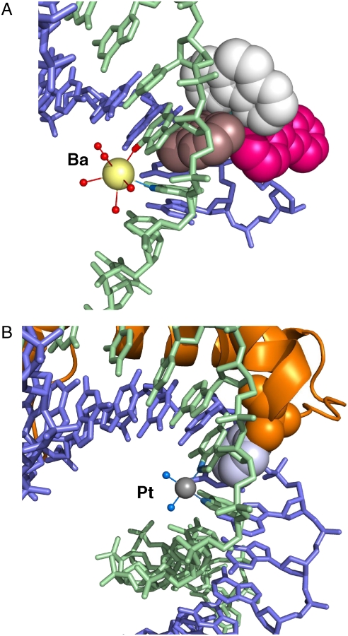
Fig. 5.
Kinking by platination in contrast to semiintercalation by 1. (A) Hydration of the barium ion and its coordination to N7 of guanine G3 and O6 of guanine G4 in the major groove. For Ba-ligand distances, see Table S2. The color scheme for 1 is as in Figs. 3A and 4. (B) Cisplatin-DNA adduct bound to high-mobility group B1 (HMGB1), Protein Data Bank ID 1CKT (37), in an approximately similar orientation. Platinum is shown as a small gray sphere, NH3 ligands as small blue spheres, HMGB1 as an orange ribbon, with the intercalating phenyl side chain of phenylalanine in space-fill mode with the phenyl group in gray. The platinum is directly coordinated to two N7 residues on adjacent guanine bases.

