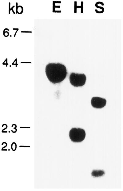
Figure 3.
Southern-blot analysis of the AtGSK1 gene. Genomic DNA was digested with restriction enzymes and size separated onto a 0.8% agarose gel. The DNA was then transferred onto a nylon membrane and UV cross-linked. Hybridization was carried out with the AtGSK1 cDNA at 65°C overnight. E, H, and S indicate EcoRI, HindIII, and SacI, respectively.

