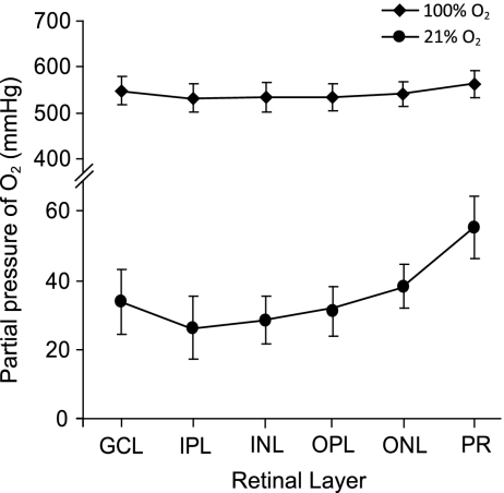
Fig. 2.
Partial pressure of O2 within the ex vivo retina exposed to 21% and 100% O2. Retinas perfused with 100% O2-bubbled saline had a much higher pO2 in all retinal layers. GCL, ganglion cell layer; IPL, inner plexiform layer; INL, inner nuclear layer; OPL, outer plexiform layer; ONL, outer nuclear layer; PR, photoreceptors.

