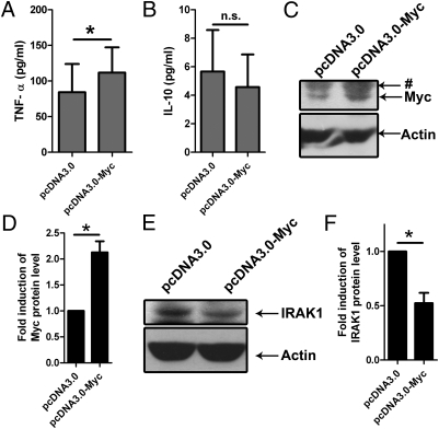
Fig. 5.
Overexpression of Myc is sufficient for TNF-α induction and IRAK1 activation. PBMac were electroporated with the indicated expression plasmid and incubated with culture medium for 48 h. Supernatants were discarded and the cells were incubated with fresh culture medium for 24 h. (A and B) Supernatants were harvested for determining the levels of the indicated cytokines by ELISA. The data are expressed as mean ± SEM from four (A) and three (B) independent blood donors. (C and E) Total proteins were harvested for analysis by Western blot. The data are representative results from cells isolated from three independent blood donors. (D and F) Protein levels of Myc (D) and IRAK1 (F) were normalized with that of Actin and expressed as fold induction relative to the cells electroporated with pcDNA3.0 plasmid. The data are expressed as mean ± SEM from three independent blood donors. The * and n.s. denote P < 0.05 and P > 0.05, respectively, as determined by Student's t test. The # denotes nonspecific bands.

