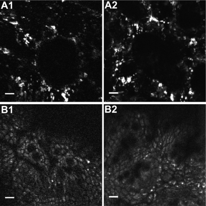
Fig. 6.
Imaging comparison between the multiphoton endoscope and a commercial multiphoton microscope. (A) Unaveraged intrinsic fluorescence images of ex vivo mouse lung. A1 shows the image acquired from the multiphoton endoscope, and A2 shows the image acquired from the Olympus multiphoton microscope. (B) Five frames averaged intrinsic fluorescence images of ex vivo mouse colon. B1 shows the image acquired from the multiphoton endoscope, and B2 shows the image acquired from the Olympus multiphoton microscope. Scale bars, 10 μm. Comparable amounts of two-photon excited fluorescence at the sample per frame were maintained in each system. The displayed images were acquired with 800-nm excitation and a FOVxy of approximately 110 μm × 110 μm.

